Overview
What is Portal Hypertension?
Portal hypertension refers to elevated blood pressure within the portal venous system, a network of veins draining abdominal organs into the portal vein, which then directs blood flow into the liver. This condition can lead to the backward flow of blood into various abdominal organs, including the spleen, esophagus, and rectum, potentially causing bleeding.
Various factors can contribute to portal hypertension, restricting blood flow through the portal vein, hepatic veins, or the liver itself. Examples of conditions leading to portal hypertension include portal vein clots, liver disease, and right heart failure, with chronic liver scarring, known as cirrhosis, being the most common cause.
Risk Factors for Portal Hypertension:
- Cirrhosis (Liver Scarring)
- Hemochromatosis
- Congestive Heart Failure
- Arteriovenous Malformations (AVMs)
- Hypercoagulable States
Symptoms of Portal Hypertension:
Symptoms may not manifest until pressure levels become high, resulting in complications like:
- Ascites (fluid in the abdomen)
- Hepatic encephalopathy (confusion)
- Variceal bleeding
- Vomiting of blood
- Bloody or black stools
- Enlarged spleen (Splenomegaly) with potential platelet decrease and left abdominal pain or fullness
Additionally, liver failure symptoms indicative of portal hypertension include jaundice, easy bruising or bleeding, weakness, weight loss, and abdominal pain.
If you have symptoms or risk factors, seek a consultation with Dr. Mohamed Hosni for accurate diagnosis and timely intervention. Early detection under his specialized care enhances the effectiveness of portal hypertension treatment.
Causes and Diagnoses of Portal Hypertension:
Causes of Portal Hypertension:
- Cirrhosis (Liver Scarring): The most prevalent cause is often linked to chronic liver diseases.
- Portal Vein Clots: Obstruction of the portal vein due to blood clots.
- Liver Disease: Various liver disorders can contribute to portal hypertension.
- Right Heart Failure: Impaired heart function affects blood flow.
Diagnoses of Portal Hypertension:
- Endoscopy: Uses a camera in the gastrointestinal tract to detect variceal bleeding.
- Abdominal Ultrasound: Imaging to visualize the abdominal organs and blood vessels.
- Hepatic Venous Pressure Gradient (HVPG): Measures the pressure within the portal system.
- CT or MRI Scans: Provides detailed images of the abdominal area.
Additional tests may include platelet level assessments, liver function tests, liver biopsy, and angiography to pinpoint the underlying causes and related conditions.
Treatments for Portal Hypertension:
Medical interventions for portal hypertension may include:
- Transjugular intrahepatic portosystemic shunt (TIPS)
- Balloon-occluded retrograde transvenous obliteration (BRTO) or percutaneous antegrade transvenous obliteration (PARTO)
Transjugular Intrahepatic Portosystemic Shunt (TIPS)
Treatment for: Ascites, Portal hypertension
Why it’s done:
Liver disease can cause a restriction of flow through the liver, leading to elevated pressure in the circulation from the bowel to the liver (portal vein). High portal vein pressures can result in bleeding (esophageal or gastric varices) and the buildup of fluid in the abdomen (ascites) or the chest (hepatic hydrothorax).
How it’s done:
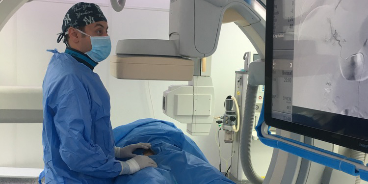
An interventional radiologist utilizes ultrasound and X-rays to guide a catheter into the vein draining the liver. A specialized needle is then passed through the inside of the liver and into the portal vein. A covered stent is placed to create a shunt from the portal vein to the hepatic vein, bypassing the liver and relieving the elevated portal vein pressure.
Tips: The procedure is performed under general anesthesia.
Risks:
In addition to the mentioned risks, TIPS may also entail the potential for stent dysfunction, development of new varices, or heart-related complications.
Post-procedure:
Most patients are admitted for several days for monitoring. Post-procedure, close monitoring is essential, and patients may experience discomfort or pain at the incision site.
Follow-up:
Doppler ultrasound is performed 2-7 days after TIPS to ensure the shunt remains open. Follow-up appointments, usually every 6 months afterward, involve imaging studies to evaluate the shunt's patency and overall liver function. These appointments are crucial for adjusting treatment plans as needed and ensuring the long-term success of the procedure.
Balloon-Occluded Retrograde Transvenous Obliteration (BRTO)
Treatment for: Gastric varices, Hepatic encephalopathy, Portal hypertension
Why it’s done:
Patients with liver disease often experience high pressure in the portal venous system. Elevated pressure can lead to the enlargement of collateral veins, often within the lining of the stomach (gastric varices) or from the spleen to the veins draining the kidney (splenorenal shunt). Gastric varices, being thin-walled and fragile, pose a risk of life-threatening bleeding. Additionally, portosystemic shunts like splenorenal shunt can allow portal venous blood to bypass the liver's filtering effect, leading to toxins reaching the brain and causing confusion (hepatic encephalopathy). Blocking these varices or shunts can eliminate the risk of bleeding and reduce confusion.
How it’s done:
An interventional radiologist initiates the procedure by accessing a vein in the neck or groin. A catheter is maneuvered into the enlarged collateral vein(s) draining the portal venous system, and the collateral veins (varices or shunts) are blocked using coils, gelfoam, or other agents.
Level of anesthesia: Conscious sedation or general anesthesia
Risks:
In addition to the mentioned risks, BRTO may also carry a risk of renal failure, thrombosis, or pulmonary embolism.
Post-procedure:
Patients are admitted for observation for 2-3 days. Post-procedure, a CT scan is typically performed to confirm the successful occlusion of varices/shunts. Monitoring for bleeding, infection, or worsening ascites is crucial during this period.
Follow-up:
Regular follow-up in the interventional radiology clinic every 6 months involves assessments to confirm continued occlusion of varices/shunts and the effective control of symptoms. This ongoing evaluation is vital for the long-term management of portal hypertension and its associated complications.
