Overview
Understanding Biliary Interventions: A Comprehensive Guide
What Is a Biliary Intervention?
Biliary interventions are minimally invasive procedures to address narrowed or blocked bile ducts. The liver produces bile, a digestive fluid that flows through ducts to the gallbladder and then to the small intestine. When these ducts are obstructed, it can lead to complications such as jaundice, nausea, and inflammation. Biliary drains, also known as stents or catheters, alleviate these issues by draining excess bile from the liver.
Symptoms of Biliary Intervention:
- Jaundice
- Belly pain
- Nausea
- Fever
- Itching
- Dark urine
- Light stools
- Lack of appetite
Types of Biliary Interventions:
Percutaneous Transhepatic Cholangiography (PTC):
Image-guided needle insertion into the liver. Contrast material injection to visualize the biliary tract. Additional procedures may include catheter insertion or stent placement.
Percutaneous Transhepatic Biliary Drainage (PTBD):
Catheter insertion into blocked liver ducts for bile drainage.
Percutaneous Cholecystostomy:
Catheter placement into an infected gallbladder for drainage.
Role of Interventional Radiologists:
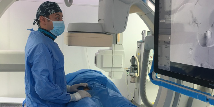
Interventional radiologists perform these procedures using imaging guidance, such as fluoroscopy, CT, and ultrasound. They employ these techniques for biopsies, fluid drainage, catheter insertion, and stent placement to address narrowed ducts and blood vessels.
Causes and Diagnoses of Biliary Interventions:
Several conditions can lead to blockages or narrowing in the bile duct. These include:
- Inflammation:
- Cholangitis (liver inflammation)
- Cholecystitis (gallbladder inflammation)
- Primary Sclerosing Cholangitis (bile duct scarring)
- Cancer:
- Pancreatic, gallbladder, bile duct, or liver cancer
- Enlarged lymph nodes from various tumors
- Gallstones: Present in the gallbladder or bile ducts
- Surgical Injury: Bile duct damage during surgery
Equipment Used in Procedures:
X-ray Equipment:
This procedure involves a radiographic table, one or two X-ray tubes, and a video monitor. Fluoroscopy, which converts X-rays into video images, is used for real-time guidance.
Ultrasound:
Ultrasound scanners consist of a computer console, a video display screen, and a transducer for body and blood vessel scans. The transducer sends high-frequency sound waves into the body, creating an image based on echoes.
CT Scanning:
A CT scanner is a large, box-like machine with a central hole. During the scan, you'll lie on a narrow table that moves in and out of the scanner. The X-ray tube and detectors rotate around you, capturing cross-sectional slices of your body.
Additional Equipment:
Catheter: A thin plastic tube used in procedures. Balloon: A thin plastic tube with a small balloon at the end. Stent: A small wire mesh or plastic tube.
Treatments: How Biliary Interventions Work
Biliary interventions typically initiate with percutaneous transhepatic cholangiography, utilizing x-rays and contrast material to visualize the bile ducts and gallbladder. When encountering a blockage, the doctor may employ various methods, including:
- Drainage Tube Placement: Draining excess bile from the body.
- Bile Duct Opening: Dilating a narrowed bile duct.
- Stent Placement: Inserting a stent to maintain bile duct openness.
- Restoring Bile Flow: Reestablishing the normal flow of bile within the biliary system.
Procedure Overview:
Percutaneous Transhepatic Cholangiography (PTC):
A thin needle is guided through the skin into the liver using ultrasound and x-ray. Contrast material injection aids in imaging the biliary tract.
Percutaneous Transhepatic Biliary Drainage (PTBD):
In case of blockage, a catheter is inserted into liver ducts for external bile drainage. Three drainage methods: external, internal/external, and internal with stent placement.
Percutaneous Cholecystostomy:
A catheter is inserted through the skin into the gallbladder using ultrasound and x-ray. Allows drainage of bile fluid into a collection bag outside the body.
Patient Experience:
Before the Procedure:
Diagnostic imaging (ultrasound, CT, or MRI) may be performed. Medications for nausea, pain, and infection prevention may be administe red.
During the Procedure:
Intravenous (IV) line insertion for sedative administration. General anesthesia or conscious sedation may be used. Small skin incision at the procedure site.
After the Procedure:
Monitoring of heart rate and blood pressure. Sensations of a slight pinch during IV line insertion and local anesthetic injection. Warmth sensation during contrast material injection.
Benefits vs. Risks:
Benefits:
- Minimal surgical incisions, often requiring no stitches.
- Shorter hospitalization compared to open surgery.
- Speedier recovery time.
Risks:
- Infection risk (1 in 1,000 cases).
- Minimal risk of allergic reaction to contrast material.
- Small risk of bleeding, often self-resolving or treatable with minimally invasive techniques.
- Potential risks associated with drainage tubes include swelling, bleeding, infection, blockage, and malposition.
Limitations of Biliary Interventions:
While minimally invasive, these procedures may not be suitable for all patients. Your doctor and interventional radiologist will determine the appropriateness based on your specific situation. Generally, minimally invasive approaches are preferable over surgery.
Prioritize your biliary health. Book an appointment with Dr. Mohamed Hosni for expert guidance and personalized care. Your journey to wellness starts here!