Liver transplantation can be done by full transplantation from the cadaver or from a living donor to obtain a lobe of the liver.
The transplant is anastomosed (connected) to the corresponding vessels in the liver, portal vein, and biliary tract recipient.
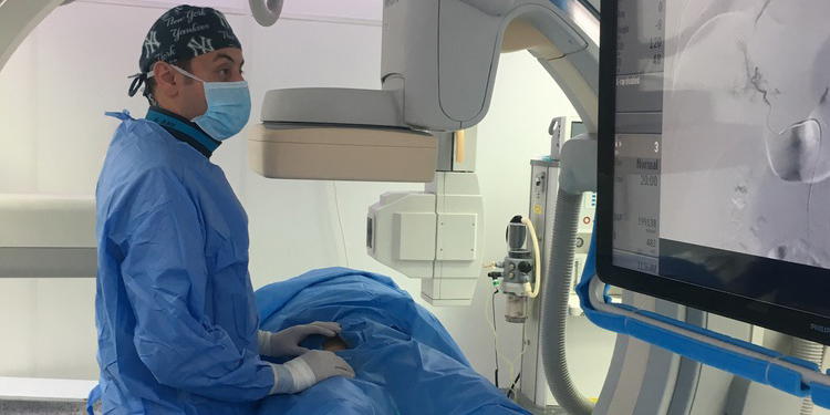
Interventional radiology is prominent in the treatment of complications arising from these anastomoses.
Complications due to arterial and venous anastomosis are more common in the early period, while it is prominent with complications due to anastomosis in the long term.
In the treatment of arterial and venous anastomosis complications, re-surgery and interventional radiological methods can be used.
Early thrombosis, rupture of the vessels (dissection), false or true vessel ballooning (aneurysm or pseudoaneurysm), stenosis can be treated with closed angiographic methods and the transplantation organ can maintain its functions.
Decrease in the flow of bile to the intestines due to stenosis that may occur in the biliary tract may lead to the development of jaundice, a biliary tract called cholangitis and severe biliary tract infection.
After liver transplants, biliary stenosis can be seen in 20-25%.
In the treatment, it is entered through the skin and passed through the liver and a thin tube is placed in the bile ducts to provide the normal flow of biliary tracts.
In case of need, patient control sessions are called to expand the stenosis areas in various parts of the biliary tract by inflating the balloon or by applying the ablation procedures that we call ablation.
In addition, melting stents may be placed in some stenosis areas.
Treatment of biliary stenosis can take several sessions.