Percutaneous Endoscopic Gastrostomy (PEG): Overview
Modern Medicine and Imaging Technologies
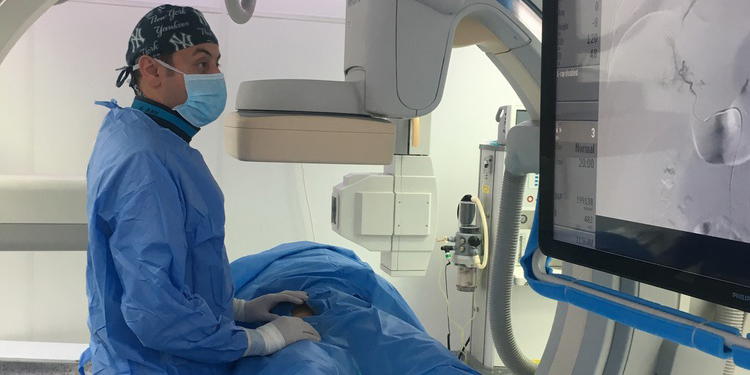
Modern medicine has been revolutionized by imaging technologies, allowing doctors to see inside the human body with unprecedented clarity. Among the pioneers of this transformation are interventional radiologists, specialists who use imaging not only to diagnose diseases but also to guide minimally invasive treatments. This field represents the forefront of precision medicine, where interventions are tailored to the specific needs of each patient. In this article, we’ll explore how interventional radiologists use imaging to save lives and improve outcomes, offering a behind-the-scenes look at this cutting-edge medical specialty.
The Role of Imaging in Interventional Radiology
Imaging lies at the heart of interventional radiology (IR). Techniques such as X-rays, computed tomography (CT), magnetic resonance imaging (MRI), and ultrasound enable interventional radiologists to visualize internal structures in real-time. These tools guide the placement of needles, catheters, and other devices with remarkable precision, minimizing risks and enhancing treatment effectiveness.
Key Imaging Modalities:
- X-Ray Fluoroscopy: Provides continuous real-time imaging, often used in procedures like angioplasty or stent placement.
- CT Scans: Offers detailed cross-sectional views of the body, essential for guiding biopsies or ablations.
- MRI: Delivers high-resolution images of soft tissues, ideal for diagnosing and treating tumors.
- Ultrasound: Utilizes sound waves to create real-time images, frequently employed for vascular access or fluid drainage.
Life-Saving Applications of Imaging in IR
Interventional radiologists use imaging to address a wide range of medical conditions, from life-threatening emergencies to chronic diseases. Here are some of the most impactful applications:
1. Treating Blood Clots and Blocked Arteries
- Thrombectomy: Interventional radiologists use imaging to guide catheters into blood vessels to remove clots directly. Techniques like mechanical thrombectomy can rapidly restore blood flow in stroke or DVT patients.
- Angioplasty and Stenting: With the help of fluoroscopy, narrowed or blocked arteries can be widened using balloons or supported with stents, restoring circulation and preventing complications like heart attacks.
2. Cancer Diagnosis and Treatment
- Biopsies: Imaging ensures precise targeting of suspicious lesions, allowing for accurate diagnosis with minimal discomfort to the patient.
- Ablation Techniques: Methods like radiofrequency ablation (RFA) and microwave ablation (MWA) use heat to destroy tumors. Imaging ensures that the treatment is focused entirely on the cancerous tissue.
- Embolization: Interventional radiologists use imaging to guide tiny particles or coils into blood vessels feeding tumors, cutting off their blood supply. Techniques like transarterial chemoembolization (TACE) and radioembolization (TARE) combine embolization with chemotherapy or radiation.
3. Emergency Interventions
- Trauma Management: Interventional radiologists use angiography to identify and control internal bleeding, often preventing the need for open surgery.
- Aortic Aneurysm Repair: Imaging guides the placement of stent grafts to reinforce weakened blood vessels, preventing rupture and saving lives.
4. Managing Chronic Conditions
- Fluid Drainage: Using ultrasound or CT guidance, interventional radiologists drain abscesses, cysts, or pleural effusions, relieving symptoms and preventing complications.
- Pain Management: Imaging-guided nerve blocks or vertebroplasty procedures provide relief for patients with chronic pain due to cancer or spinal fractures.
Precision Medicine: A Tailored Approach
Interventional radiology embodies the principles of precision medicine by tailoring treatments to the individual needs of each patient. This approach involves:
- Targeted Therapy: Imaging allows for the precise delivery of treatments, such as chemotherapy or radiation, directly to the affected area.
- Minimally Invasive Techniques: Procedures are performed through small incisions or punctures, reducing recovery times and complications.
- Real-Time Monitoring: Imaging ensures that interventions are performed accurately, with the ability to make adjustments as needed during the procedure.
The Technology Behind the Scenes
The success of interventional radiology depends on advanced imaging technologies and tools that enhance precision and safety.
Imaging Innovations:
- 3D Imaging: Provides a detailed view of anatomical structures, helping interventional radiologists plan and execute complex procedures.
- Fusion Imaging: Combines data from different imaging modalities, such as CT and MRI, to create a comprehensive view of the target area.
- Artificial Intelligence (AI): AI algorithms are being integrated into imaging systems to enhance diagnostic accuracy and guide interventions.
Navigation Tools:
- Electromagnetic Navigation Systems: Allow for precise tracking of instruments within the body, even in challenging cases.
- Robotic Assistance: Robotic systems are increasingly used to enhance the precision of needle placements and other interventions.
The Patient Experience
For patients, interventional radiology offers numerous benefits compared to traditional surgical approaches:
- Reduced Risks: Imaging-guided procedures involve smaller incisions, leading to lower risks of infection, bleeding, and other complications.
- Shorter Recovery Times: Most IR procedures are performed on an outpatient basis or require only a brief hospital stay.
- Minimal Discomfort: Local anesthesia is often sufficient, reducing the need for general anesthesia and its associated risks.
- Improved Outcomes: The precision of imaging-guided treatments often leads to better results, whether in terms of symptom relief or disease management.
Real-Life Impact: Stories of Success
Case 1: Stroke Patient
A 55-year-old man experiencing an acute ischemic stroke undergoes mechanical thrombectomy guided by real-time fluoroscopy. The clot is successfully removed, restoring blood flow to the brain and preventing permanent disability.
Case 2: Cancer Treatment
A woman with inoperable liver cancer undergoes TACE, with imaging guiding the delivery of chemotherapy directly to the tumor. The treatment shrinks the tumor significantly, extending her life expectancy and improving her quality of life.
Case 3: Trauma Intervention
After a car accident, a young man with internal bleeding receives an emergency embolization procedure. Imaging pinpoints the source of the bleed, and the intervention stops it without the need for open surgery.
Challenges and Future Directions
While interventional radiology has made remarkable strides, challenges remain:
- Access to Technology: Advanced imaging equipment can be costly, limiting its availability in some regions.
- Training and Expertise: Interventional radiologists require specialized training to perform these highly technical procedures.
The future of interventional radiology looks promising, with ongoing advancements in imaging and precision tools:
- Personalized Medicine: Integrating genetic and molecular data with imaging will enable even more tailored treatments.
- Expanded Applications: Research is exploring new uses for interventional radiology, from treating autoimmune diseases to enhancing organ transplantation.
Conclusion
Interventional radiologists are at the forefront of precision medicine, using imaging to diagnose, treat, and manage a wide range of conditions. Their expertise in minimally invasive techniques not only saves lives but also enhances the quality of care for patients. As technology continues to advance, the role of imaging in interventional radiology will only grow, offering new possibilities for saving lives and improving outcomes. Behind every successful procedure is the skilled hand of an interventional radiologist and the transformative power of imaging.
